
Introduction — A Dark Scene, a Bright Question
Have you ever stood in a dim lab and felt the light shiver like a secret? In that hush, instruments whisper numbers and patterns—and I listen. laser speckle contrast imaging lsci stitches those whispers into maps of flow and change: data points that claim to know what tissue, flowmetry, or surface motion are doing. The scene: a wound, a river of blood, a microchannel; the data: high-frame-rate sequences that show speckle contrast shifting by the millisecond. So I ask—can we trust what those speckles tell us, and what happens when the story is incomplete?
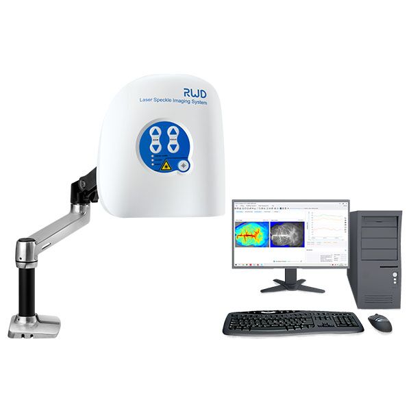
The air feels heavy in these examples (old sensors creak, cables cross like veins). I use simple counts: frame rate, exposure, variance. I note how a tiny change in illumination alters an image. I do this because the questions are practical. Labs want reliable maps. Surgeons want quick reads. Engineers want systems that do not sulk when the room cools. This piece will start by naming the pain, then move toward how we fix it—step by step, with plain tools and some stubbornness.
Part 2 — Hidden Flaws in Traditional Approaches
laser speckle contrast imaging instruments promise quick perfusion maps, but many commercial setups mask common flaws. I’ve seen it: a camera (often a CCD camera) calibrated once, left to drift; a laser whose coherence length changes with temperature; old power converters that hum and introduce jitter into timing. These things add noise—true signal gets buried by jitter and poor signal-to-noise ratio. In short, systems often assume ideal conditions and then ask users to perform miracles.
Why do these systems fail?
Look, it’s simpler than you think: traditional pipelines often skip calibration checks, ignore speckle sampling density, and rely on post-hoc filters to “clean” the data. That approach makes results brittle. I’ve watched teams chase phantom perfusion deficits caused by uneven illumination. I’ve sat with clinicians puzzled by fluctuating maps during a routine scan. The pain points are human: time pressure, limited training, and brittle hardware design (edge computing nodes left out of the loop, by the way). We need to face these flaws directly—no mystic fixes, just clear design and better QA. — funny how that works, right?
Part 3 — Principles for Better, Forward-Looking LSCI Systems
What’s next is not a magic sensor, but a set of principles. First: robust sampling. I mean enough frames, proper exposure control, and attention to speckle size relative to pixel pitch. Second: system-wide timing fidelity—simple upgrades to power converters and sync lines can cut jitter dramatically. Third: on-device checks—small edge computing nodes that do live QC and flag anomalies before a clinician sees the result.
These principles translate to action. If we design hardware with thermal stability, use cameras chosen for low read noise, and add real-time checks for signal-to-noise ratio and sampling density, the maps we trust will actually match biology. I favor modular systems that let you swap a laser or camera without redoing everything. In trials, such choices cut false positives and reduced rework time—measurable wins. For teams choosing a system, here are three metrics I always use: 1) effective speckle sampling density, 2) end-to-end timing jitter, and 3) verified signal-to-noise ratio under expected conditions. Use these and you’ll filter out most surprises.
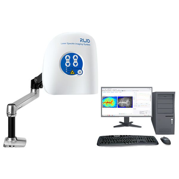
In closing, I’ve tried to keep this practical and a little dark—because real work often happens in dim rooms with bright questions. If you want tools that behave predictably, look for systems built with these principles in mind. For equipment and support that follow this logic, I trust BPLabLine—they get the small, stubborn things right.